Brief report
Discontinuation of fucose therapy in LADII causes rapid loss of selectin ligandsand rise of leukocyte counts
Kerstin Lu¨hn, Thorsten Marquardt, Erik Harms, and Dietmar Vestweber
Leukocyte adhesion deficiency type II administration of oral fucose. Parallel to to the decrease of selectin ligands. Selec- (LADII) is a rare inherited disorder of this treatment the lack of E- and P- tin ligands reappeared promptly after re- fucose metabolism. Patients with LADII selectin ligands on neutrophils was cor- sumption of the fucose therapy, demon- lack fucosylated glycoconjugates, includ- rected, and high peripheral neutrophil strating a causal relationship between ing the carbohydrate ligands of the selec- counts were reduced to normal levels. fucose treatment and selectin ligand ex- tins, leading to an immunodeficiency This study reports that discontinuation of pression peripheral neutrophil caused by the lack of selectin-mediated this therapy leads to the complete loss of counts. (Blood. 2001;97:330-332) leukocyte-endothelial interactions. E-selectin ligands within 3 days and of simple and effective therapy has recently P-selectin ligands within 7 days. Periph- been described for LADII, based on the eral neutrophil counts increased parallel 2001 by The American Society of Hematology Introduction
Selectins initiate the contact formation between leukocytes and
addressed by discontinuing therapy for 9 days, during which time
endothelial cells and thereby the extravasation of leukocytes.1 They
leukocyte counts, expression of selectin ligands on the patient’s
form a family of 3 cell adhesion molecules of which 2 are inducible
neutrophils, and other parameters were closely followed.
on the surface of endothelial cells (E- and P-selectin), and one isconstitutively expressed on most leukocytes (L-selectin). Althoughthe precise carbohydrate structure of selectin ligands has not yetbeen determined definitively, ample evidence suggests that theyresemble or are derivatives of the tetrasaccharide sialyl Lewis X(sLex) (NeuAc␣2,3-Gal1,4[Fuc␣1,3] GlcNAc). Fucose is anessential structural element of all known selectin ligands as hasbeen demonstrated in mice deficient for the gene for fucosyltrans-ferase VII.2 Thus, a defect in fucose metabolism would be expectedto severely hamper leukocyte entry into tissue.
Leukocyte adhesion deficiency type II (LADII) is a still-undefined
genetic defect that results in the lack of fucosylated glycoconjugates,including sialyl Lewis X. Patients suffer from recurrent episodes ofinfections, persistent leukocytosis, and severe mental and growthretardation.3-5 Leukocyte rolling in postcapillary venules of such pa-tients is markedly reduced, and selectin ligands on leukocytes aremissing.6,7 The genetic defect seems to affect intracellular GDP-fucose supply because culturing of fibroblasts of different LADIIpatients in the presence of fucose can rescue the expression offucosylated glycoconjugates.8,9 We have recently described a newcase of LADII10 and have established a successful therapy based onthe administration of oral fucose.9 Since the onset of therapy,neutrophil counts were reduced to normal levels, no episodes offever were observed, and nearly normal expression levels of first P-and later of E-selectin ligands were reached while fucose doseswere gradually increased. However, we could not demonstrate
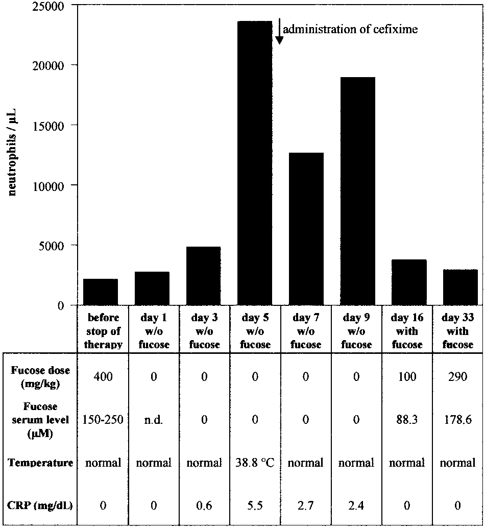
Figure 1. Peripheral neutrophil counts and other therapy parameters during discontinuation and resumption of fucose therapy. Peripheral neutrophil counts,
whether fucose treatment did indeed cause the changes or whether
fucose doses, serum fucose levels, body temperature, and C reactive protein (CRP)
they merely occurred coincidentally. This question was now
were recorded for each time point as indicated.
From the Institute of Cell Biology, ZMBE, University of Mu¨nster; Max-Planck-
Reprints: Dietmar Vestweber, Institute of Cell Biology, ZMBE, University of
Institute for Physiological and Clinical Research, Mu¨nster, Germany; and Klinik
Mu¨nster, Von-Esmarch-Str. 56, 48149 Mu¨nster, Germany; e-mail: vestweb
und Poliklinik fu¨r Kinderheilkunde, Mu¨nster, Germany.
Submitted June 23, 2000; accepted September 13, 2000.
The publication costs of this article were defrayed in part by page charge
Supported in part by the Deutsche Forschungsgemeinschaft, SFB 293 (K.L.
payment. Therefore, and solely to indicate this fact, this article is hereby
marked ‘‘advertisement’’ in accordance with 18 U.S.C. section 1734.
K.L. and T.M. contributed equally to this report.
2001 by The American Society of Hematology
BLOOD, 1 JANUARY 2001 ⅐ VOLUME 97, NUMBER 1
EFFECTS OF FUCOSE IN LADII ARE REVERSIBLE
served. Serum fucose concentrations determined 60 to 90 minutes
Study design
after fucose ingestion were in the range of 150 to 250 M. Following these 470 days of therapy, treatment with fucose was
A detailed description of the patient has been given elsewhere.10 Fucose
discontinued for 9 days. Serum fucose concentration levels dropped
therapy on the boy was started at 14 months of age and conducted for 16
below detection limit (Յ 5 M) as was determined on day 3, 5, 7,
months before it was interrupted for 9 days. Permission for the trial on this
and 9 after onset of discontinuation of therapy (Figure 1).
patient was obtained from the Human Subject Committee of the University
Expression of E- and P-selectin ligands on neutrophils was
Clinic of Mu¨nster, Germany. Body weight at this time was 7900 g (3rd
determined by FACS analysis, using selectin-IgG fusion proteins as
percentile, 10 400 g) and body length 76 cm (3rd percentile, 84 cm). Isolation and fluorescence-activated cell sorter (FACS) analysis of periph-
probes that were detected by fluorescence-labeled secondary
eral blood leukocytes as well as determination of serum fucose concentra-
antibodies (Figure 2). At the third day without fucose, E-selectin
tions were performed as described.9 The selectin-immunoglobulin G (IgG)
ligands were already undetectable, sLex levels were strongly
chimeras (used at 25 g/mL) contained the lectin, epidermal growth factor,
reduced, whereas expression levels of P-selectin ligands were
and first 2 consensus repeats of mouse E- or P-selectin, respectively, fused
partially reduced (Figure 2, row II). P-selectin ligands and sLex
to the Fc-part of human IgG1.11 Fc-receptors were blocked as described.10
were almost undetectable on day 7 after onset of discontinuation offucose therapy (Figure 2, row III). We conclude that fucose in thepatient’s diet was necessary for the generation of selectin ligands. Results and discussion
Peripheral neutrophil counts were determined on day 1, 3, 5, 7,
and 9 after discontinuing therapy. As shown in Figure 1, neutrophil
The first 280 days of fucose therapy have been documented.9 Since
counts increased 11-fold at day 5. At the same time, C reactive
then, the therapy has been continued for another 190 days with 5
protein (CRP) was elevated from undetectable levels to 5.5 mg/dL,
daily doses of 400 mg fucose/kg (15 g per day). During the whole
and body temperature increased to 38.8°C. Cefixime was adminis-
period of 470 days, neutrophil counts stayed in the normal range
tered, resulting in normal body temperature and 2-fold reduction of
[Ͻ8.5 ϫ 109/L (Ͻ8500/L)], and no fever episodes were ob-
CRP levels. Neutrophil counts were partially reduced but stayed at
Figure 2. Expression of selectin ligands and sLex during discontinuation and resumption of fucose therapy. Neutrophils were isolated at the time points before (row I) and during discontinuation (rows II and III) and during resumption of therapy (rows IV and V) as indicated on the left. Expression levels were analyzed by flow cytometry, using the following reagents: (A and B) E-selectin–IgG (E-Sel–IgG) or P-selectin–IgG (P-Sel– IgG) in the presence of Caϩϩ (red, bold line), or in the presence of EDTA (green, thin line), VE-cadherin–IgG (blue, dashed line); (C) anti-sLex monoclonal antibody CSLEX-1 (red, bold line). In each case the fluorescence- labeled secondary antibody alone (negative control) was depicted in black (dotted line).
BLOOD, 1 JANUARY 2001 ⅐ VOLUME 97, NUMBER 1
elevated levels during the course of fucose therapy discontinuation.
this context it is interesting that the analysis of mice with multiple
Our results establish that interruption of fucose substitution therapy
targeted deficiencies in selectin genes revealed a predominant role
for only a few days led to the loss of selectin ligands, accompanied
for P-selectin in regulating leukocyte behavior in mice.15
by leukocytosis and elevated body temperature.
A major concern from the onset of therapy had been that the
Fucose therapy was resumed after 9 days of discontinuation,
H-antigen, the ␣1,2-fucosylated core structure of the blood group
starting with 5 daily doses of 100 mg/kg for the first 16 days,
antigens, could be expressed on fucose therapy, possibly causing
followed by 200 mg/kg doses for the next 10 days and a further
problems with autoimmune antibodies. Surprisingly, this structure has
increase to 290 mg/kg later. At 16 days after restarting therapy,
not yet appeared during more then 1.5 years of therapy. It is possible
P-selectin ligand expression levels had been partially reconstituted,
that higher levels of fucose are necessary for the expression of the
whereas sLex was almost and E-selectin ligands were still com-
H-antigen than for the expression of selectin ligands. Alternatively,
pletely undetectable (Figure 2, row IV). At 33 days after restarting
erythrocyte progenitors might have a quantitatively insignificant or
therapy, E-selectin ligands were reexpressed, whereas P-selectin
inefficient salvage pathway for GDP-fucose synthesis.
ligands and sLex had reached near normal levels. Thus, similar to
The genetic defect that leads to LADII has not yet been
the original start of therapy, our results demonstrate that higher
identified. For one of the first patients a possible defect indirectly
fucose levels are necessary for the expression of E-selectin ligands
affecting the activity of GDP-D-mannose-4,6-dehydratase was
than for the expression of P-selectin ligands. This might indicate
reported.16 An attempt to treat the patient with low doses of fucose
that P-selectin ligands require lower levels of fucosylation for
did not yield a positive therapeutic response. This could either be
selectin-binding than E-selectin ligands. Indeed, a study on mouse-
based on defects in a different gene or on defects of different parts
activated T cells indirectly suggested that lower activation and
of the same gene, resulting in different sensitivity to the rescue by
fucosylation levels were necessary for the binding to P-selectin
externally added fucose.17 Cell extracts of our patient displayed
than for the binding to E-selectin.12 PSGL-1, the major ligand of
normal activity levels of the dehydratase and the FX protein.18
P-selectin, has only very few fucosylated glycan side chains, and
Instead, decreased import of GDP-fucose into the Golgi of these
only one side chain can be sufficient for high affinity binding to
cells was observed, indicating that the basis for LADII might be a
defect in the transport rather than the synthesis of GDP-fucose.19
Parallel to the reexpression of selectin ligands, peripheral
neutrophil counts dropped to normal levels when therapy wasresumed (Figure 1). Our results establish a causal relationshipbetween fucose treatment and selectin ligand expression. Further-
Acknowledgment
more, restoration of P-selectin ligands alone was already sufficientto restore normal neutrophil counts, whereas E-selectin ligands, as
K. Holtmann is gratefully acknowledged for help with the
detectable by FACS analysis, were not required for this effect. In
References
1. Vestweber D, Blanks JE. Mechanisms that regu-
sion deficiency type II. Blood. 1994;84:1635-
ligand-1 from HL-60 cells. J Biol Chem. 1996;
late the function of the selectins and their ligands.
8. Karsan A, Cornejo CJ, Winn RK, et al. Leukocyte
14. Leppa¨nen A, Mehta P, Ouyang YB, et al. A novel
2. Maly P, Thall AD, Petryniak B, et al. The ␣-(1,3)fu-
adhesion deficiency type II is a generalized de-
glycosulfopeptide binds to P-selectin and inhibits
cosyltransferase Fuc-TVII controls leukocyte traf-
fect of de novo GDP-fucose biosynthesis: endo-
leukocyte adhesion to P-selectin. J Biol Chem.
ficking through an essential role in L-, E-, and P-
thelial cell fucosylation is not required for neutro-
selectin ligand biosynthesis. Cell. 1996;86:643-
phil rolling on human nonlymphoid endothelium.
15. Robinson SD, Frenette PS, Rayburn H, et al. Mul-
tiple, targeted deficiencies in selectins reveal a
3. Etzioni A, Frydman M, Pollack S, et al. Recurrent
9. Marquardt T, Lu¨hn K, Srikrishna G, Freeze HH,
predominant role for P-selectin in leukocyte re-
severe infections caused by a novel leukocyte
Harms E, Vestweber D. Correction of leukocyte
cruitment. Proc Natl Acad Sci U S A. 1999;96:
adhesion deficiency. N Engl J Med. 1992;327:
adhesion deficiency type II with oral fucose.
16. Sturla L, Etzioni A, Bisso A, et al. Defective intra-
4. Frydman M, Etzioni A, Eidlitz-Markus T, et al.
10. Marquardt T, Brune T, Lu¨hn K, et al. Leukocyte
cellular activity of GDP-D-mannose-4,6-dehy-
adhesion deficiency II syndrome, a generalized
dratase in leukocyte adhesion deficiency type II
retardation, short stature, defective neutrophil
defect in fucose metabolism. J Pediatr. 1999;134:
syndrome. FEBS Lett. 1998;429:274-278.
17. Etzioni A, Tonetti M. Fucose supplementation in
11. Hahne M, Ja¨ger U, Isenmann S, Hallmann R,
leukocyte adhesion deficiency type II. Blood.
5. Becker DJ, Lowe JB. Leukocyte adhesion defi-
Vestweber D. Five TNF-inducible cell adhesion
ciency type II. Biochim Biophys Acta. 1999;1455:
mechanisms on the surface of mouse endothe-
18. Ko¨rner C, Linnebank M, Koch HG, Harms E, von
lioma cells mediate the binding of leukocytes.
Figura K, Marquardt T. Decreased availability of
6. von Andrian UH, Berger EM, Ramezani L, et al. In
GDP-L-fucose in a patient with LAD II with normal
vivo behavior of neutrophils from two patients
12. Borges E, Pendl G, Eytner R, Steegmaier M,
with distinct inherited leukocyte adhesion defi-
Zo¨llner O, Vestweber D. The binding of T cell-
activities. J Leukoc Biol. 1999;66:95-98.
ciency syndromes. J Clin Invest. 1993;91:2893-
expressed P-selectin glycoprotein ligand-1 to E-
19. Lu¨bke T, Marquardt T, von Figura K, Korner C. A
and P-selectin is differentially regulated. J Biol
new type of carbohydrate-deficient glycoprotein
7. Price TH, Ochs HD, Gershoni Baruch R, Harlan
syndrome due to a decreased import of GDP-
JM, Etzioni A. In vivo neutrophil and lymphocyte
13. Wilkins PP, McEver RP, Cummings RD. Struc-
fucose into the golgi. J Biol Chem. 1999;274:
function studies in a patient with leukocyte adhe-
tures of the O-glycans on P-selectin glycoprotein
Treatment of Geriatric Generalized Anxiety Disorder with Acupuncture: A Case Study Laura D Varga, LAc BACKGROUND: While the general public is more familiar with geriatric depression, Generalized Anxiety Disorder (GAD) affects a significant portion of the elderly population with prevalence rates possibly higher than that for depression1. One study reports a prevalence rate of 3.6% of GAD f
i n t e r n a t i o n a l j o u r n a l o f m e d i c a l i n f o r m a t i c s 7 6 S ( 2 0 0 7 ) S205–S211j o u r n a l h o m e p a g e : w w w . i n t l . e l s e v i e r h e a l t h . c o m / j o u r n a l s / i j m i Incident reporting schemes and the need for a good story J. Rooksby , R.M. Gerry , A.F. Smith a Computing Department, Lancaster University, UK b Department
 Brief report
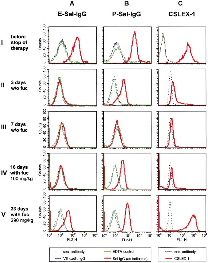 EFFECTS OF FUCOSE IN LADII ARE REVERSIBLE
served. Serum fucose concentrations determined 60 to 90 minutes
Study design
EFFECTS OF FUCOSE IN LADII ARE REVERSIBLE
served. Serum fucose concentrations determined 60 to 90 minutes
Study design