Nephrol Dial Transplant (2003) 18: 54–61
Estradiol is nephroprotective in the rat remnant kidney
Balazs Antus1, Peter Hamar1, Gabor Kokeny1, Zoltan Szollosi2, Istvan Mucsi1,3, Zoltan Nemes2and Laszlo Rosivall1
1Department of Pathophysiology, Semmelweis University, Budapest, Hungary, 2Department of Pathology, MedicalUniversity, Debrecen, Hungary and 3First Department of Internal Medicine, Semmelweis University, Budapest, Hungary
female sex hormones, such as estrogens, may be
Background. Female sex hormones may influence
responsible for the lower susceptibility of women to
the progression of renal diseases. We therefore eva-
luated the effects of estradiol on the development of
Glomerulosclerosis and atherosclerosis may share
glomerulosclerosis in a remnant kidney model.
common elements in their pathogenesis. It has been
Methods. Ovariectomized or intact female Wistar rats
suggested that the renoprotective effects of estrogens
underwent 5u6 nephrectomy. Ovariectomized animals
may be related to their effects on glomerular mesangial
were treated with vehicle, 17b-estradiol alone or in
cells in a manner analogous to the effects of estrogens
combination with progesterone, intact rats received
on vascular smooth muscle cells in atherosclerosis.
vehicle only. Twenty-four weeks after renal ablation,
In support of this hypothesis, estradiol has been
histological as well as molecular analysis were
shown to suppress cellular proliferation as well as the
synthesis of type I and IV collagen, and to inhibit trans-
Results. Vehicle-treated ovariectomized animals deve-
forming growth factor (TGF)-b- anduor platelet-
loped severe proteinuria and glomerulosclerosis as
derived growth factor (PDGF)-mediated type IV
compared with vehicle-treated intact rats. In addition,
collagen expression in mesangial cells [2]. Further-
renal mRNA levels of platelet-derived growth factor-A
more, estradiol induces the synthesis of matrix metallo-
chain (PDGF-A) were increased. Estradiol replace-
proteinases in mesangial cells, suggesting that estrogens
ment reduced proteinuria, which was paralleled by a
may limit glomerular scarring by increasing matrix
diminished glomerular injury and reduced transform-
ing growth factor-b1 (TGF-b1) and PDGF-A mRNA
The renoprotective properties of estrogens, however,
expression. In animals that received combined hor-
have been recently challenged. Baylis et al. [4] reported
mone treatment there were no significant differences in
that age-related glomerulopathy in female rats is not
proteinuria, creatinine clearance, renal histopathology
influenced by estrogens. Similarly, Mulroney et al. [5]
and growth factor mRNA levels compared with those
demonstrated that ovariectomy has no effect on early
measured in vehicle-treated ovariectomized rats. Serum
development of glomerular hypertrophy and glomer-
cholesterol and triglyceride levels were comparable
ular injury in uninephrectomized female rats. Finally,
between all groups during the whole follow-up period.
in obese Zucker rats and in Nagase analbuminemic
Conclusions. The data suggest that estrogens protect
rats, administration of estradiol impairs renal function
against the development of glomerulosclerosis in the
and induces profound glomerulosclerosis [6,7].
It has been argued that co-administration of pro-
gesterone with estrogens can modulate the cardio- and
Keywords: estradiol; glomerulosclerosis; growth factors;
atheroprotective effects of estrogens. Some investiga-
tors have observed a decrease in the beneficial effectsof estrogens [8], while other studies demonstrated noadverse effects of progesterone addition [9]. Never-
theless, little information is available as to whether theaddition of progesterone to estrogens influences the
Progression of chronic renal failure is slower in women
effects of estrogens in renal injury.
than in men. Recently, it has been speculated that
Thus, in the present study we investigated the role
of estrogens in the development of progressiveglomerulosclerosis after subtotal renal ablation in
Correspondence and offprint requests to: Dr Balazs Antus, Semmelweis
female Wistar rats. Furthermore, we tested whether
University, Department of Pathophysiology, Nagyvarad ter
H-1089 Budapest, Hungary. Email: antbal@net.sote.hu
co-administration of progesterone with estrogens
# 2003 European Renal Association–European Dialysis and Transplant Association
modulates the effects of estrogens on this process.
pressure (Hemosys, Experimetria, Hungary) was measured.
Finally, we studied whether growth factors such as
Thereafter, rats were bled and the kidneys and the uteri
TGF-b and PDGF, which are commonly associated
were removed. Remnant kidneys were cut into two pieces.
with progressive glomerulosclerosis, are affected by
One kidney sample was snap frozen in liquid nitrogen formolecular analysis, the other piece was fixed in buffered
Fixed kidney tissues were embedded in paraffin and stained
using haematoxylinueosin, periodic acid–Schiff (PAS) and
Seven-week-old female Wistar rats (Charles River, Hungary)
Masson’s trichrome methods. PAS reaction was performed
weighing 160–200 g were used in these experiments. Animals
to evaluate the extent of glomerulosclerosis. Glomerulosclerosis
were kept under standard conditions and given free access
was defined as the accumulation of extracellular matrix in
to tap water, and the same amount of standard rat chow
the mesangium. Collapse of capillaries and adhesion of
obsolescent segments of Bowman’s capsule were frequently
uweekuanimal). All experiments were approved by a
governmental committee on animal welfare.
seen in the sclerosed glomeruli. Glomerulosclerosis wasevaluated according to the following scoring method: score0, normal glomerulus; score 1, mild segmental glomerulo-
sclerosis affecting -25% of the glomerular tuft; score 2,moderate segmental glomerulosclerosis affecting 25 to 50%
All rats were subjected to subtotal (5u6) renal ablation under
of the glomerular tuft; score 3, diffuse severe glomerulo-
sodium-pentobarbital (55 mgukg, i.p.) anaesthesia as des-
sclerosis affecting )50% of the glomerular tuft. A mini-
cribed previously [10]. Briefly, after removal of the right
mum of 40 glomeruli per remnant kidney was examined,
kidney, the left kidney was subtotally resected by removing
and the mean of the glomerular scores was taken to
from the cortex two-thirds of the weight of the resected right
represent the severity of glomerulosclerosis for a given rat.
kidney. Special care was taken to avoid damage to pelvis
The degree of tubulointerstitial fibrosis (tubulointerstitial
and hilum. To stop bleeding, renal vessels of the left kidney
damage index) was evaluated in trichrom-stained sections
were clamped for 5 min in all operated animals including
and graded according to the following scale: 0, no evidence
ovariectomized and intact animals. The excised kidney tissue
of interstitial fibrosis; grade 1, lesions involving -25% of
was weighed on an analytic scale. The average reduction of
the tubulointerstitial area; grade 2, lesions affecting 25–
the total kidney mass was 74 " 1.4%.
involving )50% of the tubulointerstitial area. All histo-pathological evaluations were carried out by two indepen-
dent observers (B. Antus and Z. Szollosi) blinded to the
Following renal ablation a total number of 32 animals were
randomly allocated into the following four experimentalgroups (ns8ugroup) according to gonadal status and hor-monal treatment: animals in the first group remained intact
and received vehicle (INT). In the second, third and fourth
Total RNA was extracted with Trizol (GibcouBRL, Life
groups, rats were ovariectomized and treated with either 17b-
Technologies, Germany) according to the protocol provided
estradiol (E), 17b-estradiolqprogesterone (EqP) or vehicle
by the manufacturer. Briefly, frozen tissues were mixed with
(OVX). 17b-Estradiol (20 mgukg; Sigma, Sigma Aldrich,
1 ml Trizol reagent, homogenized, mixed with 0.2 ml chloro-
Germany) and progesterone (10 mgukg, Sigma) were dis-
form, and centrifuged at 12 000 g for 15 min at 48C. RNA,
solved in sesame oil and administered subcutaneusly every
from the aqueous phase, was precipitated with 0.5 ml
second day (0.1 ml) until harvesting, similarly to our pre-
isopropyl alcohol and centrifuged at 12 000 g for 10 min.
vious studies [11]. Vehicle-treated intact (INT) and ovar-
iectomized (OVX) animals were given sesame oil alone. The
centrifuged at 7500 g for 5 min and dried. RNA was
reduction of the renal mass was similar between the groups
dissolved in DEPC-treated water and stored at À808C.
(INT, 73.7 " 3.7%; OVX, 75.2 " 1.8%; E, 74.1 " 2.6% andEqP, 73.1 " 3.1%).
Reverse transcriptase–polymerase chain reaction
RNA was amplified by reverse transcription (RT) with anOligo(dT)12–18 primer (Perkin-Elmer, Applied Biosystem,
Every 4 weeks, body weights were measured and 24-h
Germany) using 1 mg of total RNA added to 0.5 mg of
urine samples were collected using metabolic cages and a
primer. The reaction mixture contained: buffer solution
urine-cooling system. Urine protein was determined nephelo-
[TRIS hydrochloride (50 mM, pH 8.3); potassium chloride
metrically, while serum cholesterol and triglyceride concen-
trations were determined using a Reflotron analyzer
(5 mM)], adenosine triphosphate, thymidine triphosphate,
guanosine triphosphate and cytosine triphosphate each at a
At week 24, serum as well as urine creatinine levels were
concentration of 0.2 mM (GibcouBRL), 0.5 ml of 40 Uuml
measured to determine creatinine clearance. Furthermore,
of recombinant ribonuclease inhibitor (Perkin-Elmer) and
serum 17b-estradiol and progesterone concentrations were
0.5 ml of 200 Uuml M-MLV reverse transcriptase (GibcouBRL).
measured by radioimmunassay using commercially available
The reaction was allowed to proceed (428C, 1 h), then it
kits (Immunotech, Izinta, Hungary). Animals were then
was halted by heating the samples to 958C for 5 min
Specific cDNA products corresponding to mRNA for
weight, as well as the uterus weight-to-body weight ratio,
TGF-b1, PDGF-A chain and b-actin were amplified using
was significantly lower in vehicle-treated ovariecto-
polymerase chain reaction (PCR) as described previously
mized (OVX) animals as compared to vehicle-treated
[10,11]. Briefly, 1 ml from RT reaction was taken for PCR,
intact rats (INT) (Table 1). Estrogen replacement
which was performed in PCR buffer [750 mM Tris–HCl,
alone (E) or in combination with progesterone (EqP)
pH 9.0, 200 mM (NH4)2SO4, 0.1% (wuv) Tween 20, 20 mM
maintained uterus weight similar to animals with
magnesium dichloride (Qiagen, Germany)] using 0.2 mM of
each deoxynucleoside triphosphates, 1 mM of both primersand 2.5 U thermus Aquaticus (Taq) DNA polymerase(Qiagen). A Perkin-Elmer Thermal Cycler (Model 9600,Perkin-Elmer, Norwalk, CT) was used for amplification with
the following sequence profile: initial denaturation at 948C
By week 24, vehicle-treated ovariectomized animals
for 3 min followed by 30–35 cycles (denaturing: 948C for
developed increased proteinuria as compared to
30 s; annealing: 558C for 30 s; extension: 728C for 30 s) and
ending with a final extension at 728C for 7 min.
versus INT 283.2 " 47.5 mgu24 h, P-0.05, ANOVA)
The amplified PCR products were identified by electro-
(Figure 1). Furthermore, serum creatinine levels were
phoresis of 10 ml aliquots on 1.5% agarose gel stained with0.5 mg
elevated and there was a trend towards a decreased
uml of ethidium bromide. Specific products were
visualized by UV transillumination and identified by size in
creatinine clearance in these rats (Table 2). Estradiol
relation to a 1 kb oligonucleotide DNA ladder (GibcouBRL).
replacement reduced both proteinuria (E, 286.4 " 39.1
Intensities of the specific bands were semiquantitated by
versus OVX, 556.6 " 112.4 mgu24 h, P-0.05, ANOVA)
densitometric analysis, and the ratios of the density of the
and serum creatinine to approximately the same level
specific bands to the bands of b-actin (internal control) were
as in vehicle-treated intact rats. Similarly, the creati-
nine clearance was better maintained in estradiol-treatedanimals as compared to vehicle-treated ovariectomized
rats. Co-administration of progesterone with estradioltended to reduce the beneficial effects of estradiol both on
Data are presented as mean " SEM. Parametric data were
urinary protein excretion (E, 286.4 " 39.1 mgu24 h
compared using one-way analysis of variance (ANOVA),
versus EqP, 383.6 " 62.6 mgu24 h) and serum crea-
followed by multiple pair-wise comparison according to the
tinine, but these differences did not reach statistical
Newman–Keuls test. Non-parametric data were tested using
Kruskal–Wallis one-way analysis of ranks. A P value of-0.05 was considered significant.
Ovariectomy elicited a significant reduction of
plasma 17b-estradiol levels (Table 2). Estradiol levelsin rats treated with estradiol alone (E) or in combina-
tion of progesterone (EqP) were within the rangefor intact female rats reported in the literature [12]and were not significantly different from values for
At the beginning of this study, body weights were
throughout the estrus cycle) in the present study. One
comparable between the groups (Table 1). At week 24,
estradiol-treated rat with extremely high estradiol
body weight was significantly higher in vehicle-treated
levels ()300 pguml) was excluded from this study.
ovariectomized (OVX) rats compared to intact (INT)
Ovariectomy also reduced serum progesterone con-
or sex hormone-treated animals (E, EqP). Further-
centrations (Table 2). Addition of progesterone to
more, sex hormone-treated animals tended to have a
estradiol treatment (EqP) resulted in plasma concentra-
decreased body weight compared with intact rats.
tions of progesterone similar to those reported previously
Atrophy of the uterus is considered to be a sensitive
in the literature [12] or obtained in vehicle-treated
indicator of the completeness of ovariectomy. Uterus
intact animals (INT) in the present study.
Table 1. Body and uterus weight at the time of renal ablation and after 24 weeks
aP-0.005 vs vehicle-treated intact animals (ANOVA).
bP-0.01 vs vehicle-treated intact animals (ANOVA).
cP-0.01 vs vehicle-treated ovariectomized animals (ANOVA).
Fig. 1. Changes in 24-h urinary protein excretion throughout the study. (*P-0.05 vs vehicle-treated intact animals; §P-0.05 vsvehicle-treated ovariectomized animals).
Table 2. Mean arterial blood pressure and serum values
aP-0.05 vs vehicle-treated intact animals (ANOVA).
bP-0.01 vs vehicle-treated ovariectomized animals (ANOVA).
cP-0.01 vs vehicle-treated intact animals (ANOVA).
dP-0.005 vs 17b-estradiol-treated ovariectomized animals (ANOVA).
Mean arterial blood pressure seemed to be the
diminished glomerular injury in these animals was
highest in vehicle-treated ovariectomized animals, but
accompanied by a significantly lower degree of
the difference between the groups was not significant
(Table 2). Similarly, lipid levels, both serum choles-
Both glomerulosclerosis index and the degree of
terol and triglyceride were comparable between the
tubulointerstitial fibrosis were elevated in animals
groups during the whole follow-up period (Figure 2A
that received the combined sex hormone replace-
and B). However, by week 24, cholesterol levels in all
ment (EqP) as compared with those given estradiol
treatment alone (E); however, these differences werenot statistical significant (Table 3). Similarly, neitherglomerulosclerosis nor tubulointerstitial fibrosis dif-
fered significantly between animals that received the
Glomerulosclerosis was significantly increased in ovar-
combined hormone replacement (EqP) or vehicle
iectomized vehicle-treated rats (OVX) as compared to
intact rats (INT) (Table 3). Similarly, we noted asignificantly higher degree of tubulointerstitial fibrosis
Glomerulosclerosis was significantly reduced in
estradiol-treated animals (E) compared to vehicle-
sion detected by semiquantitative PCR paralleled
treated ovariectomized rats (OVX) (Table 3). The
development of renal fibrosis (Figure 3). Accordingly,
ovariectomy induced an ;2-fold increase in expres-sion of PDGF-A in vehicle-treated animals (OVX,2.1 " 0.3 vs INT, 0.9 " 0.1, P-0.01, ANOVA). TGF-b1 mRNA levels were also increased in vehicle-treatedovariectomized
1.4 " 0.2), however, these differences were not sig-nificant. Estradiol replacement reduced expression ofboth TGF-b1 (OVX, 2.7 " 0.4 vs E, 1.1 " 0.2,P-0.05, ANOVA) and PDGF-A (OVX, 2.1 " 0.3vs E, 0.6 " 0.1, P-0.005, ANOVA). Animals thatreceived estradiol in combination with progesteronetended to have elevated TGF-b1 (EqP, 1.5 " 0.5 vs E,1.1 " 0.2) and PDGF-A (EqP, 1.6 " 0.3 vs E,0.6 " 0.1) expression compared to estradiol-treated rats.
In this study we found that estrogen status plays animportant role in the progression of glomerulosclerosisafter subtotal renal ablation in female rats. Estrogendeficiency in gonadectomized females was associatedwith a rapid loss of renal function that was preventedby estradiol replacement. The data suggest thatestrogens are renoprotective in a model of chronicrenal injury.
Depending on the experimental setting, estrogens
may exert various and even opposite effects on pro-gression of renal disease. In female hypercholester-olemic Imai rats, ovariectomy has been shown toaccelerate renal injury, while estradiol replacementattenuated this response [13]. Similarly, we demon-strated previously that estrogens ameliorate chronicallograft nephropathy in transplanted rat kidneys [11]. However, there are reports in contradiction with thesefindings. For example, Baylis et al. [4] reported thatage-related glomerulopathy is not influenced by estro-gens. It is possible that different mechanisms may beinvolved in glomerulopathy due to age compared withrenal ablation. For instance, glomerular hypertensionis known to play a central pathogenic role in theremnant kidney model, while this risk factor may beless important for age-dependent glomerular injury [4]. Development of compensatory renal growth and glo-
Fig. 2. (A) Changes in serum cholesterol levels throughout the study.
merular damage in uninephrectomized female rats also
(B) Changes in serum triglyceride levels throughout the study.
seems to be independent of estrogens [5]. However,
Table 3. Histological characteristics of the remnant kidneys
aP-0.05 vs vehicle-treated intact animals (Kruskal–Wallis test).
bP-0.05 vs vehicle-treated ovariectomized animals (Kruskal–Wallis test).
Fig. 3. TGF-b1 and PDGF-A chain mRNA expression in the remnant kidneys at the end of the study. (*P-0.05 vs vehicle-treated intactanimals; §P-0.05 vs vehicle-treated ovariectomized animals.)
renal function is relatively well preserved after unine-
to the more pronounced renal injury in ovariectomized
phrectomy, thus conclusions concerning the role of
rats. It is unclear whether this tendency in blood
estrogens in a progressive fibrotic process cannot be
pressure was due to the effects of estrogens, or was
drawn from those experiments. Finally, in rat strains
simply associated with the more pronounced renal
with spontaneous hypertriglyceridaemia, estrogens pro-
failure in these rats. Furthermore, it should be noted
mote renal injury [6,7]. In these models, administra-
that in our experiment blood pressure was measured in
tion of estrogens causes further increases in lipid levels,
anaesthetized rats. It is possible that the differences in
which led to progressive renal injury. Wistar rats,
blood pressure between the groups would have reached
used in our study, are normolipidaemic and neither
statistical significance, if blood pressure would have
gonadal status nor estradiol treatment influenced lipid
been measured in awake animals, for example, with
parameters. Our data, therefore, do not support a
telemetry. This technology, however, was not available
role for lipids in the renal effects of estrogens.
It is well established that dietary protein intake
Serum creatinine levels paralleled the marked histo-
influences the development of renal disease. In our
logical changes both in vehicle- and in estradiol-treated
study, animals were offered the same amount of food
animals. In contrast, creatinine clearance did not show
during the whole follow-up. Therefore, differences in
similar clear correlation with the histology. The reason
renal injury between the groups cannot be due to
for this discrepancy is not clear. However, creatinine
various protein intake. Nevertheless, vehicle-treated
clearance may be a somewhat inaccurate estimate of
ovariectomized rats gained more body weight than
renal function in rats due to the relatively high tubular
vehicle-treated intact or all sex hormone-treated rats.
creatinine secretion. This may explain why differences
Therefore, one may argue that nephronubody weight
in creatinine clearance were not statistically significant
mismatch is present and that may have contributed to
the more marked renal damage in vehicle-treated
The mechanism, by which progesterone tended to
ovariectomized rats. However, since these animals
reduce the beneficial effects of estrogen, is unclear.
accumulate mostly adipose tissue, but not muscle
Progestins are known to exert partial androgenic
mass, nephronubody weight mismatch may have had
effects weakly binding to androgen receptors in various
little effect on renal injury. Using a pair-feeding
tissues, including the kidney [14]. As androgens
protocol would have maintained body weight equal
promote renal fibrosis [4,11], it is feasible that
between the groups. However, in this case, protein
activation of androgen receptors are responsible for
consumption would have been different between the
the effects of progesterone. Furthermore, interactions
groups. Since we believe that in the reported setting
between progesterone and estrogen or progesterone
protein intake could have been a more important
and angiotensin type I receptor expression [15] could
confounder than nephronubody weight mismatch, we
controlled protein intake, but not body weight in our
Glomerular injury in the rat remnant kidney model
has been generally attributed to an altered regulation
As shown in Table 2, arterial blood pressure tended
of matrix turnover by mesangial cells. Our data are in
to be higher in vehicle-treated ovariectomized rats as
accordance with previous findings demonstrating that
compared to vehicle-treated intact animals. This
estradiol may directly limit glomerulosclerosis by
somewhat higher blood pressure may have contributed
either inhibiting collagen synthesis or increasing the
production of matrix metalloproteinases in mesangial
be higher than therapeutic doses in humans. The
reason for this difference is not clear, but variances
Furthermore, a wide range of growth factors,
between therapeutic doses in animal and human
including TGF-b1 and PDGF have been implicated
studies may perhaps be associated with differences in
in the development of glomerulosclerosis [16]. TGF-b1
promotes matrix synthesis and inhibits its degradation
In conclusion, we demonstrated that estradiol has a
by several mechanisms, and is therefore one of the
protective effect on the kidney during progressive
most important mediators of tissue fibrosis [17].
glomerulosclerosis in the female rat remnant kidney
Similarly, PDGF, apart from its strong mitogenic
model. Further studies are clearly needed to assess the
properties, stimulates production of various compo-
potential applicability of these experimental findings in
nents of extracellular matrix in human and experi-
mental renal diseases [11,18]. Moreover, it has recentlybeen suggested that estrogens may have a direct
atheroprotective effect through inhibition of TGF-b1
Research Foundations (OTKA 29260, 34409, F034498, ETT232
and PDGF-A expression in vascular smooth muscle
and FKFP 316) and the Hungarian Kidney Foundation. Dr Mucsi
cells [19]. As mesangial cells are phenotypically similar
is a recipient of the Be´ke´sy Postdoctoral Scholarship from the
to smooth muscle cells, we hypothesized that the
Hungarian Ministry of Education, Dr. Hamar is a Bolyai ResearchFellow of the Hungarian Academy of Sciences. The skilful
renoprotective effects of estradiol may be mediated, at
assistance of Maria Godo at the Department of Pathophysiology,
least in part, by its inhibitory effects on growth factor
Semmelweis University and Agnes Kovacs at the 2nd Department
synthesis in mesangial cells. Indeed, estradiol treatment
of Internal Medicine, Semmelweis University was gratefully
down-regulated the increased TGF-b1 and PDGF-A
expression after ovariectomy, and this may havecontributed to a better outcome in this group. Theco-administration of progesterone, however, tended to
reduce the beneficial effects of estradiol on expressionof TGF-b1 and PDGF-A that may be responsible for
1. Silbiger SR, Neugarten J. The impact of gender on the
progression of chronic renal disease. Am J Kidney Dis 1995;
the more pronounced glomerular injury in the animals
receiving combined hormone treatment. TGF-b1 and
2. Lei J, Silbiger S, Ziyadeh FN, Neugarten J. Serum stimulated a1
PDGF-A are important mediators of tissue fibrosis in
type IV collagen gene transcription is mediated by TGFb and
both the glomeruli and in the tubulointerstitial space.
inhibited by estradiol. Am J Physiol 1998; 274: F252–258
In this work we did not distinguish whether the
3. Potier M, Elliot SJ, Tack I, Lenz O, Striker GE, Striker LJ, Karl
observed changes in the mRNA expression reflect
M. Expression and regulation of estrogen receptors in mesangialcells: influence on matrix metalloproteinase-9. J Am Soc Nephrol
glomerular or interstitial changes in the remnant
kidneys. However, since estradiol reduced both glo-
4. Baylis C. Age-dependent glomerular damage in the rat.
merulosclerosis and interstitial fibrosis, we assume that
Dissociation between glomerular injury and both glomerular
TGF-b and PDGF expression were down-regulated
hypertension and hypertrophy. Male gender as a primer risk
parallel in both compartments of the kidney by the
factor. J Clin Invest 1994; 94: 1823–1829
5. Mulroney SE, Woda C, Johnson M, Pesce C. Gender-differences
There are insufficient data available at present to
in renal growth and function after uninephrectomy in adult rats. Kidney Int 1999; 56: 944–953
determine whether estrogen replacement therapy can
6. Gades MD, Sern JS, van Goor H, Nguyen D, Johnson PR,
influence progression of renal disease in postmeno-
Kaysen GA. Estrogen accelerates the development of renal
pausal women. This issue, however, is of considerable
disease in female Zucker rats. Kidney Int 1998; 53: 130–135
importance since the number of postmenopausal
7. Joles JA, van Goor H, Koomans HA. Estrogen induces
women with end-stage kidney disease has increased
glomerulosclerosis in analbuminemic rats. Kidney Int 1998;
sharply in the past decade. Our results suggest that
8. Hanke H, Hanke S, Bruck B et al. Inhibition of the
estrogen replacement can retard the decline in renal
atheroprotective effects estrogens by progesterone in experi-
function in this population of women. Importantly,
mental atherosclerosis. Atherosclerosis. 1996; 121: 129–138
Szekacs et al. [20] have recently demonstrated, in
9. Adams MR, Kaplan JR, Manuck SB et al. Inhibition of
a small number of diabetic and hypertensive post-
coronary artery atherosclerosis by 17b-estradiol in ovariecto-
menopausal women, that hormone replacement ther-
mized monkeys. Lack of effects of added progesterone.
apy reduced proteinuria and improved creatinine
10. Hamar P, Peti-Peterdi J, Szabo A et al. Interleukin-2-dependent
mechanisms are involved in the development of glomerulo-
Doses of sex hormones administered in our experi-
sclerosis after partial renal ablation in rats. Exp Nephrol 2001;
ment were determined in a pilot study. In those
experiments we observed that estradiol and progester-
11. Mu¨ller V, Szabo A, Viklicky O, Gaul I, Po¨rtl S, Philipp T,
one levels in sera increase up to 4 and 12 h after drug
Heemann U. Sex hormones and gender related differences: their
administration with doses that we applied in our
influence on chronic renal allograft rejection. Kidney Int 1999;55: 2011–2020
present experiments, and then they return to the base
12. The control of progesterone secretion during the estrous cycle
line 48 h later. These hormone levels were comparable
and early pseudopregnancy in the rat: prolactin, gonadotropin
with those in naive cycling rats. Of note is the fact that
and steroid levels associated with rescue of the corpus luteum of
steroid doses administered in our experiment seem to
pseudopregnancy. Endocrinology 1975; 96: 219–226
13. Sakemi T, Toyoshima H, Shouno Y, Morito F. Estrogen
17. Yamamoto T, Noble NA, Miller DE, Border WA. Sustained
attenuates progressive glomerular injury in hypercholesterolemic
expression of TGF-b1 underlies development of progressive
male Imai rats. Nephron 1995; 69: 159–165
kidney fibrosis. Kidney Int 1994; 45: 916–927
14. Bullock LP, Bardin CW, Sherman MR. Androgenic, antiandro-
18. Floege J, Burns MW, Alpers CE. Glomerular cell proliferation
genic and synandrogenic actions of progestins: role of steric and
and PDGF expression precede glomerulosclerosis in the remnant
allosteric interactions with androgen receptors. Endocrinology
kidney model. Kidney Int 1992; 41: 297–309
19. Kikuchi N, Urabe M, Iwasa K et al. Atheroprotective effect of
15. Nickening G, Strehlow K, Wassmann S et al. Differential
estradiol and estrone sulfate on human vascular smooth muscle
effects pf estrogen and progesterone on AT1 receptor geneexpression in vascular smooth muscle cells. Circulation 2000;
cells. J Steroid Biochem Mol Biol 2000; 72: 71–78
20. Szekacs B, Vajo Z, Varbiro Sz et al. Postmenopausal hormone
16. Muchaneta-Kubara EC, Sayed-Ahmed N, El Nahas AM.
replacement improves proteinuria and impaired creatinine
Subtotal nephrectomy: a mosaic of growth factors. Nephrol
clearance in type 2 diabetes mellitus and hypertension. BJOG
Received for publication: 7.1.02Accepted in revised form: 20.8.02
Name __________________________________________________ Date ____________ Date of birth _____________________ Date of last eye exam ______________________Referring Dr. ______________________________ Primary Care Physician __________________________What is the chief complaint regarding your eyes?_______________________________________________________________________________________________
The Journal of DermatologyVol. 32: 972–975, 2005A Case of Lichenoid Drug Eruption Associated Emiliano Antiga, Lucilla Melani, Carla Cardinali, Barbara Giomi, Marzia Caproni, Stefano Francalanci And Paolo Fabbri Abstract A 53-year-old man developed lichenoid lesions on the upper chest, posterior surfaces ofthe trunk, and abdominal region about three months before his first visit. Physical
 Nephrol Dial Transplant (2003) 18: 54–61
Estradiol is nephroprotective in the rat remnant kidney
Balazs Antus1, Peter Hamar1, Gabor Kokeny1, Zoltan Szollosi2, Istvan Mucsi1,3, Zoltan Nemes2and Laszlo Rosivall1
1Department of Pathophysiology, Semmelweis University, Budapest, Hungary, 2Department of Pathology, MedicalUniversity, Debrecen, Hungary and 3First Department of Internal Medicine, Semmelweis University, Budapest, Hungary
female sex hormones, such as estrogens, may be
Background. Female sex hormones may influence
responsible for the lower susceptibility of women to
the progression of renal diseases. We therefore eva-
luated the effects of estradiol on the development of
Glomerulosclerosis and atherosclerosis may share
glomerulosclerosis in a remnant kidney model.
Nephrol Dial Transplant (2003) 18: 54–61
Estradiol is nephroprotective in the rat remnant kidney
Balazs Antus1, Peter Hamar1, Gabor Kokeny1, Zoltan Szollosi2, Istvan Mucsi1,3, Zoltan Nemes2and Laszlo Rosivall1
1Department of Pathophysiology, Semmelweis University, Budapest, Hungary, 2Department of Pathology, MedicalUniversity, Debrecen, Hungary and 3First Department of Internal Medicine, Semmelweis University, Budapest, Hungary
female sex hormones, such as estrogens, may be
Background. Female sex hormones may influence
responsible for the lower susceptibility of women to
the progression of renal diseases. We therefore eva-
luated the effects of estradiol on the development of
Glomerulosclerosis and atherosclerosis may share
glomerulosclerosis in a remnant kidney model.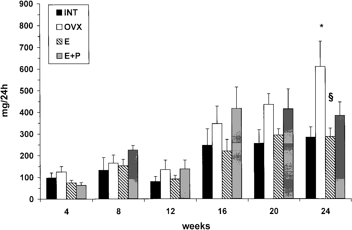 Fig. 1. Changes in 24-h urinary protein excretion throughout the study. (*P-0.05 vs vehicle-treated intact animals; §P-0.05 vsvehicle-treated ovariectomized animals).
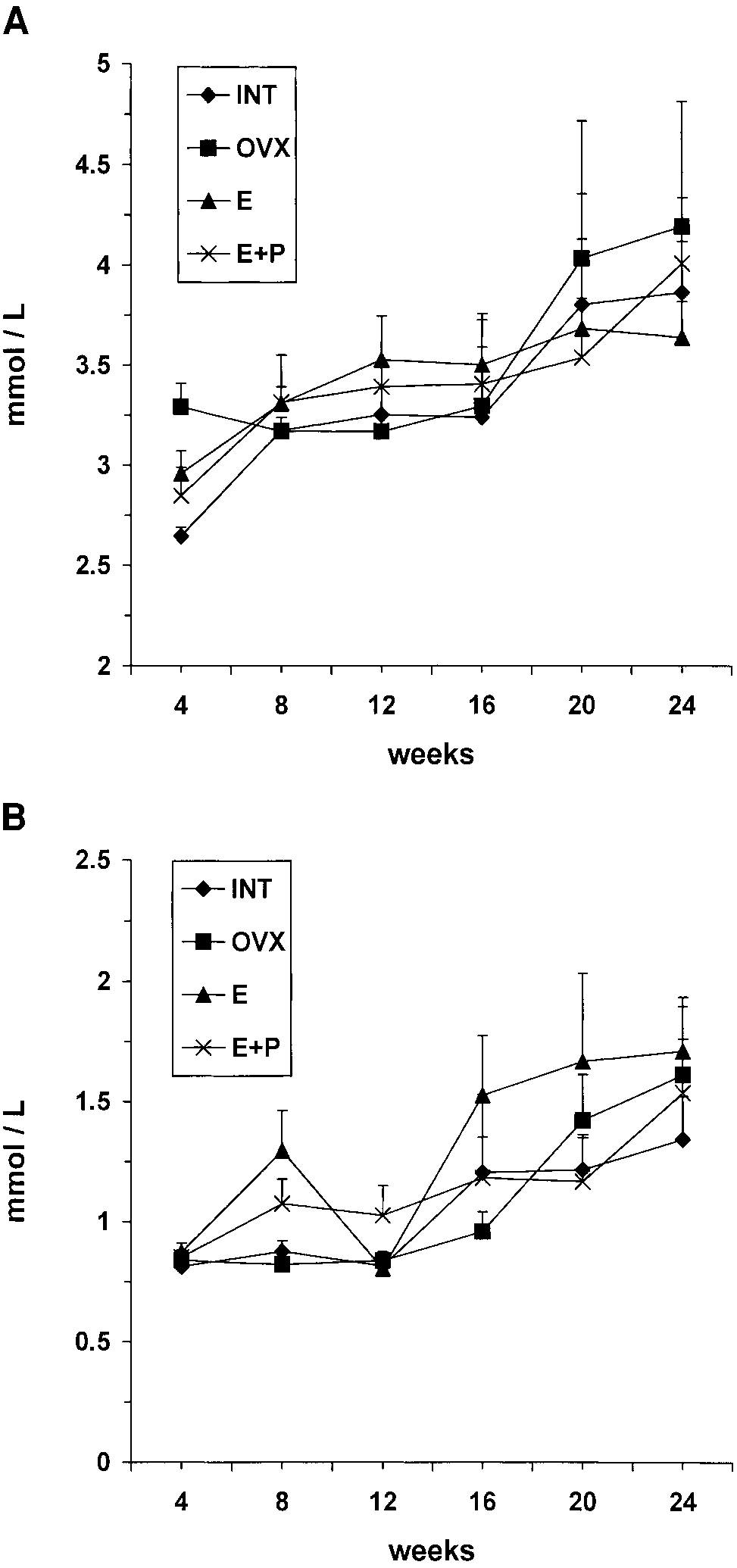 ovariectomy induced an ;2-fold increase in expres-sion of PDGF-A in vehicle-treated animals (OVX,2.1 " 0.3 vs INT, 0.9 " 0.1, P-0.01, ANOVA). TGF-b1 mRNA levels were also increased in vehicle-treatedovariectomized
1.4 " 0.2), however, these differences were not sig-nificant. Estradiol replacement reduced expression ofboth TGF-b1 (OVX, 2.7 " 0.4 vs E, 1.1 " 0.2,P-0.05, ANOVA) and PDGF-A (OVX, 2.1 " 0.3vs E, 0.6 " 0.1, P-0.005, ANOVA). Animals thatreceived estradiol in combination with progesteronetended to have elevated TGF-b1 (EqP, 1.5 " 0.5 vs E,1.1 " 0.2) and PDGF-A (EqP, 1.6 " 0.3 vs E,0.6 " 0.1) expression compared to estradiol-treated rats.
ovariectomy induced an ;2-fold increase in expres-sion of PDGF-A in vehicle-treated animals (OVX,2.1 " 0.3 vs INT, 0.9 " 0.1, P-0.01, ANOVA). TGF-b1 mRNA levels were also increased in vehicle-treatedovariectomized
1.4 " 0.2), however, these differences were not sig-nificant. Estradiol replacement reduced expression ofboth TGF-b1 (OVX, 2.7 " 0.4 vs E, 1.1 " 0.2,P-0.05, ANOVA) and PDGF-A (OVX, 2.1 " 0.3vs E, 0.6 " 0.1, P-0.005, ANOVA). Animals thatreceived estradiol in combination with progesteronetended to have elevated TGF-b1 (EqP, 1.5 " 0.5 vs E,1.1 " 0.2) and PDGF-A (EqP, 1.6 " 0.3 vs E,0.6 " 0.1) expression compared to estradiol-treated rats.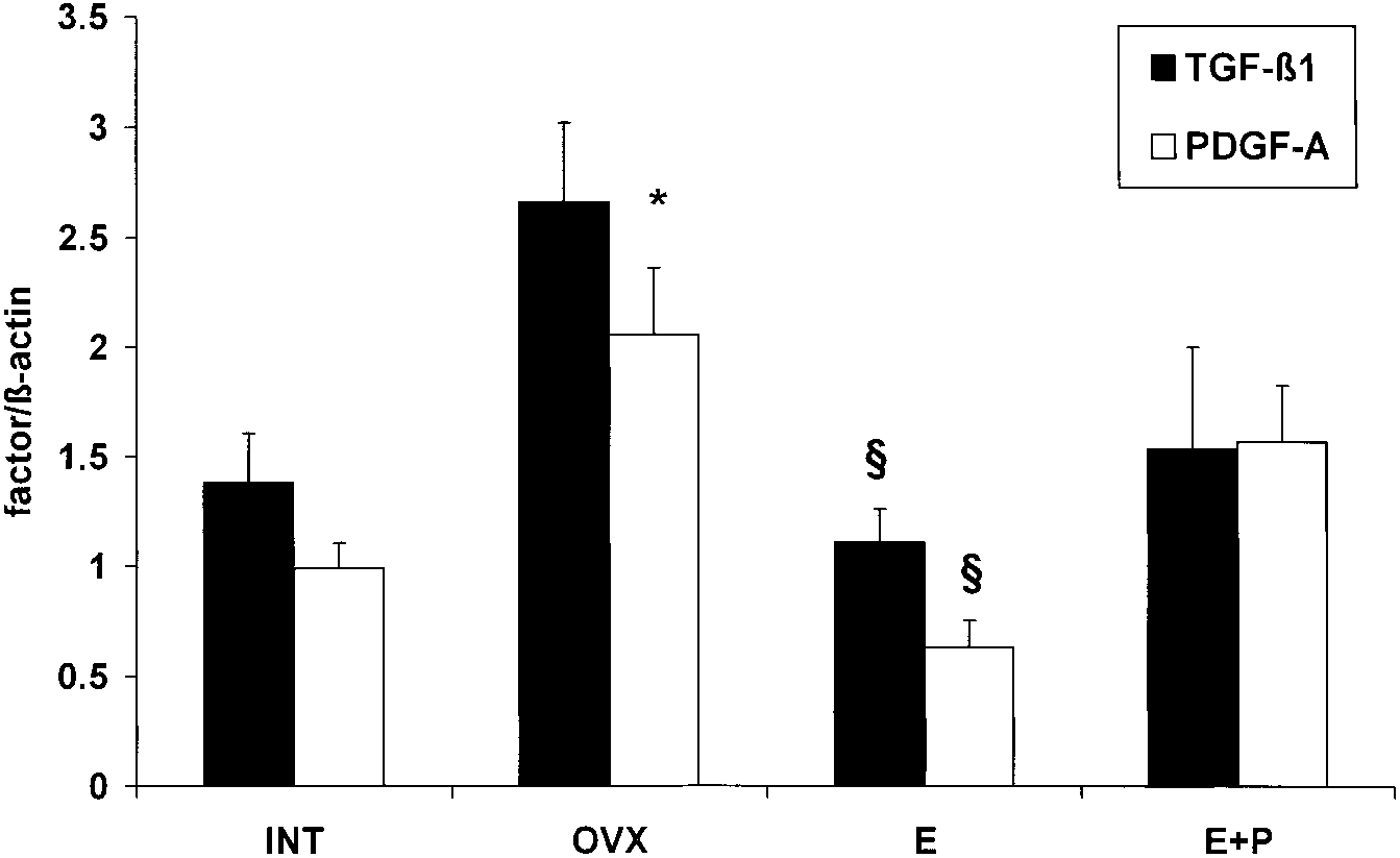 Fig. 3. TGF-b1 and PDGF-A chain mRNA expression in the remnant kidneys at the end of the study. (*P-0.05 vs vehicle-treated intactanimals; §P-0.05 vs vehicle-treated ovariectomized animals.)
renal function is relatively well preserved after unine-
to the more pronounced renal injury in ovariectomized
phrectomy, thus conclusions concerning the role of
rats. It is unclear whether this tendency in blood
estrogens in a progressive fibrotic process cannot be
pressure was due to the effects of estrogens, or was
drawn from those experiments. Finally, in rat strains
simply associated with the more pronounced renal
with spontaneous hypertriglyceridaemia, estrogens pro-
failure in these rats. Furthermore, it should be noted
mote renal injury [6,7]. In these models, administra-
that in our experiment blood pressure was measured in
tion of estrogens causes further increases in lipid levels,
anaesthetized rats. It is possible that the differences in
which led to progressive renal injury. Wistar rats,
blood pressure between the groups would have reached
used in our study, are normolipidaemic and neither
statistical significance, if blood pressure would have
gonadal status nor estradiol treatment influenced lipid
been measured in awake animals, for example, with
parameters. Our data, therefore, do not support a
telemetry. This technology, however, was not available
role for lipids in the renal effects of estrogens.
Fig. 3. TGF-b1 and PDGF-A chain mRNA expression in the remnant kidneys at the end of the study. (*P-0.05 vs vehicle-treated intactanimals; §P-0.05 vs vehicle-treated ovariectomized animals.)
renal function is relatively well preserved after unine-
to the more pronounced renal injury in ovariectomized
phrectomy, thus conclusions concerning the role of
rats. It is unclear whether this tendency in blood
estrogens in a progressive fibrotic process cannot be
pressure was due to the effects of estrogens, or was
drawn from those experiments. Finally, in rat strains
simply associated with the more pronounced renal
with spontaneous hypertriglyceridaemia, estrogens pro-
failure in these rats. Furthermore, it should be noted
mote renal injury [6,7]. In these models, administra-
that in our experiment blood pressure was measured in
tion of estrogens causes further increases in lipid levels,
anaesthetized rats. It is possible that the differences in
which led to progressive renal injury. Wistar rats,
blood pressure between the groups would have reached
used in our study, are normolipidaemic and neither
statistical significance, if blood pressure would have
gonadal status nor estradiol treatment influenced lipid
been measured in awake animals, for example, with
parameters. Our data, therefore, do not support a
telemetry. This technology, however, was not available
role for lipids in the renal effects of estrogens.