Colchicine Site Competitive Assay Kit CytoDYNAMIX Screen 15 ORDERING INFORMATION To order by phone: To order by Fax: To order by e-mail: Technical assistance: Visit the web page: CytoDYNAMIX Screen 15 Colchicine Site Competitive Assay Kit. (Cat. # CDS15) Table of contents
Introduction
Assay characterization and Unit definition
Trouble shooting and information for first time users.
CytoDYNAMIX Screen 15 Colchicine Site Competitive Assay Kit in 96-well format (Cat. # CDS15) Introduction CytoDYNAMIX Screen™ 15 is designed to detect and measure a compound’s affinity for the colchicine binding site of tubulin. The colchicine binding site is an important modulator of tubulin function, when molecules bind at this site there is inhibition of microtubule polymerization which in many cases creates a favorable therapeutic index. Examples of clinically active compounds that bind to this site are, colchicine (anti-gout), nocodazole (anti-cancer), mebendazole (anti-helminthics), T607 (anti-cancer) and benomyl (anti-fungal). More information concerning the colchicine site characteristics can be found in Hastie et al. 1989 (JBC 264, p6682-6688) and Iwasaki 1993 (Medicinal Research Reviews 13, 183-189). CDS15 can be used as a primary or secondary screen. It is economical when compared to the standard microtubule polymerization assay but it is limited by being restricting to the location of colchicines site. CDS15 assay is based on a publication by Tahir et al. 2000 (Biotechniques 29, p156-160). CDS15 is based on Scintillation Proximity Assay (SPA) technology provided by Amersham Bioscience Inc. As such this kit contains the non-proprietary items such as biotin tubulin, buffers, positive and negative controls and protocols. Items not included in the kit are plates, SPA beads and tritiated radio-ligand. These extra items are listed under the Materials and Equipment Required section. SPA technology requires a close association between a solid phase scintillant (the beads) and the radio-ligand for a signal to be emitted and subsequently detected. As shown in Figure 1, biotin tubulin is the reagent that brings the radio-ligand and the scintillant into close association. If the radio-ligand is tritiated colchicine then the amplitude of signal is proportional to the number of colchicine binding sites that are occupied by this radio ligand. When you add a competitor of radio-labeled colchicines, for example cold (unlabeled) colchicine, to the mixture then the amplitude of signal will be decreased proportionately with concentration of competitor. In some cases the inhibition can be transitory, for example with nocodazole, the reason being that colchicine binds more tightly than nocodazole but also more slowly than nocodazole, so over a long time frame (>45min) nocodazole is effectively replaced by colchicine. Figure 1: SPA assay with Biotin Tubulin and Streptavidin beads.
Kit Contents 1. 2 x 500ug
Biotin Tubulin (Cat# T333, store at -70C).
GTP Solution (100mM, BST06-001, store at -70C).
General Tubulin Buffer (BST01-010, store at 4C).
Biotin Tubulin Material (Cat.# T333) Material
Bovine brain tubulin has been modified so that random surface lysines can contain a co-
valently linked, long - chain biotin derivative. An activated ester of the biotin derivative is used in the labeling procedure. A long - chain biotin derivative was selected for this procedure because it allows the biotin molecules to be spaced far enough away from the tubulin protein so as not to interfere with subsequent visualization techniques, e.g., visualization with streptavidin based reagents. Biotin - labeled tubulin is supplied as a lyophilized powder. The powder is present usually at the bottom of the tube, but occasionally it may move to another location, follow the reconstitution instructions carefully in order to not lose the protein when opening the tube. Purity Purity is determined by scanning densitometry of proteins on SDS-PAGE gels. Samples are ≥99% pure. Biotinylation Labeling stoichiometry was determined to be approximately one biotin per tubulin heterodimer (see Figure 1A). Polymerization of T333 To test the biological activity of biotin tubulin it is polymerized in an absorbance spectrophotometer and compared to a similar unlabeled sample. Biotin tubulin has polymerization properties that are indistinguishable from those of unlabeled tubulin (see Figure 1B). Polymerizaton conditions are given in the legend to Figure 1B. Working with heterodimeric (unpolymerized) T333 If tubulin is diluted in G-PEM to below 2 mg/ml and kept at 4°C it will be maintained as tubulin heterodimers. It should be noted that tubulin is a labile protein and it should be used as soon as possible after reconstitution. Storage Figure 1 - Detection of biotin tubulin and biological activity
Figure 1: A = 10 and 100ng of T333 detected with streptavidin/alkaline phosphatase. B = Biological activity of biotin tubulin ; As determined by Microtubule assembly characteristics.Method: 1mg of biotin tubulin were reconstituted to 5 mg/ml in G-PEM buffer pH6.8 plus 5% glycerol and incubated at 35°C in a cuvette. The optical density at 340 nm was taken at time intervals. A value of 1.0 OD unit indicates that >97% of the tubulin has polymerized.
Assay Characterization and Unit Definition Example results Figure 2: Dose response with cold colchicine using the standard format.
Unit Definition Unit definition: One unit of Biotin Tubulin (Cat# T333) in CytoDYNAMIX Screen 15 is defined as 1ug of protein (as determined by the Precision Red Advanced Protein Assay, cat# ADV02). When one unit of reconstituted tubulin attached to yttrium streptavidin SPA beads is pipetted into a well containing 200ul final volume of G-PEM and 0.5uCi of tritiated colchicine at 37C, it will develop a signal of approximately 600-3000cpm depending on the sensitivity of the scintillation plate reader.
Material and equipment required Material Item Supplier Item
New England Nuclear Inc. Tel: 1-800-323-5891
Amersham Bioscience Inc. Tel: 1-800-526-3593
Not essential for CDS15 Tritiated Paclitaxel (CDS17)
American Radiolabeled Chemical Inc. Tel: 1-800-331-6661 ART1195
American Radiolabeled Chemical Inc. Tel: 1-800-331-6661 ART1194
Different batches of tritiated compounds are not made to similar specific radioactivity, batches may vary up to 5 fold in activity, it is essential to obtain a batch that has a specific activity of greater than 5Ci/mmole. Some molecules reach a theoretical maximum specific activity of 10-20Ci/mmole because the process of tritiation involves the number of acid dissociable hydrogen ions. Vinblastine for example only has only one acid dissociable hydrogen ion so its specific activity cannot be higher than 20Ci/mmole. Colchicine has several replaceable hydrogen ions so it can reach a specific activity of about 50-80Ci/mmole. Likewise paclitaxel can reach 50-80Ci/mmole. The practical implications are that the vinblastine competition assay has a much lower signal to noise ratio than the colchicine set-up. A good signal to noise with the colchicine set-up is 5:1 to 8:1 versus background, whereas vinblastine is 2:1 for 10Ci/mmole and 1:1 for 5Ci/mmole . For assay applications CDS16 (vinblastine kit) is still useful for HTS applications where a 50% difference is adequate, whereas the Ki determination is more problematic, requiring triplicate/quadruple assays for significant results. Fortunately CDS15 reagents are more economical that other tubulin assays which makes the replicate assays economically feasible. Equipment Scintillation Counter
Packard Instrument Inc. Tel: 1-800-762-4000 Topcount Microtplate Reader
Associated products: CytoDYNAMIX Screen 01 Tubulin Polymerization HTS (Absorbance, Brain, 96-well format)
CytoDYNAMIX Screen 03 Tubulin Polymerization Assay (Absorbance, Brain, 96-well format)
CytoDYNAMIX Screen 10 Hela Cell Line Tubulin Polymerization assay HTS (Fluorescence, Brain, 384-well format)
CytoDYNAMIX Screen 14 Microtubule depolymerization HTS (Fluorescence, Brain, 96 or 384-well format)
CytoDYNAMIX Screen 16 Vinblastine binding site HTS (Scintillation Proximity, 96-well format)
CytoDYNAMIX Screen 17 Paclitaxel binding site HTS (Scintillation Proximity, 96-well format)
CytoDYNAMIX Screen 18 FtsZ Polymerization
Methods: CytoDYNAMIX Screen™ 15 Introduction The assay procedure is straight forward if the set-up protocols are performed exactly as described. There are aliquots of biotin tubulin and GTP to be made as these are labile components that require -70C storage for increased stability and shelf life, which leads to improved reproducibility. As an alternative biotin tubulin is available as 20ug aliquots (T333-A or T333-B) which can be used one vial at a time for small numbers of assays. Colchicine binding is sensitive to temperature so it is important to perform the assay at 37C in a reproducible manner. The basic approach to the assay is as follows:
1. Set-up scintillation plate reader and warm plate in reader to 37°C (CDS15 only). 2. Prepare compounds to 20x strength (1mM) in G-PEM buffer plus 10% DMSO. 3. Prepare tritiated colchicine. Assay life 24h. 4. Prepare biotin tubulin on ice. Assay life 4h (four hours). 5. Prepare streptavidin beads. Assay life 24h. 6. Prepare bead/tubulin mixture. Assay life 24h. 7. Pipette 10ul of compound into the wells. 8. Pipette 10ul of tritiated colchicine into the wells. 9. Pipette 180ul of bead/tubulin into the wells. 10. Incubate for 45min at 37C. 11. Read plate. 12. Extract the raw data and calculate percent inhibition. .
The aliquots of tubulin in this kit are designed for 50 assays per run, this is useful for set-up purposes, if you require more assays per run or you require a larger assay volume because of the format of your compounds, please inquire about other aliquot sizes using tservice@cytoskeleton.com or call 303-322-2254. Reconstituted tubulin is stable for 4h in the General Tubulin Buffer plus GTP, so you can estimate the amount of tubulin required per run based on this time frame and the knowledge of the through put of your particular HTS set-up. Cytoskeleton Inc. can make any aliquot size of tubulin using it’s proprietary lyophilized tubulin bead format (Patented) please inquire at tservice@cytoskeleton.com for more information. To determine Ki’s perform a serial dilution of your compound in 3x dilutions so that you cover a range of 0.1 to 100uM using 0.1, 0.3, 1.0, 3.0, 10, 30 and 100uM and perform the assays in triplicate. Plot the Log10 of concentration versus the percent inhibition, and use the 50% inhibition intercept to read off the Ki. Tritiated compounds are not made to a constant specific radioactivity, batches may vary up to 5 fold in activity, it is essential to obtain a batch that has a specific activity of greater than 5Ci/mmole. Some molecules reach a theoretical maximum specific activity of 10-20Ci/mmole because the process of tritiation involves the number of acid dissociable hydrogen ions. Vinblastine for example only has only one acid dissociable hydrogen ion so its specific activity cannot be higher than 20Ci/mmole. Colchicine has several replaceable hydrogen ions so it can reach a specific activity of about 50-80Ci/mmole. The practical implications are that the vinblastine competition assay has a much lower signal to noise ratio than the colchicine set-up. A good signal to noise with the colchicine set-up is 5-8 fold background, whereas vinblastine is 2:1 for 10Ci/mmole and only 1:1 for 5Ci/mmole. For assay applications, CDS16 (vinblastine kit) is still useful for HTS applications where a 50% cutoff difference can be used to determine hits. The Ki determination is more problematic, requiring triplicate/quadruple assays for significant results. Fortunately CDS15 reagents are more economical that other tubulin assays which makes the multiple assays economically feasible.
Scintillation Counter Plate Reader Set-up. The majority of the work in the design of this assay has been based on a Packard Instuments Inc. machine called Topcount Microplate Scintillation Counter. This machine is one of the more sensitive machines on the market (fmoles of tritiated colchicine can be detected). The parameters of a Protocol file in this scenario are: Measurement
The major caveats with other plate readers are: 1. Low sensitivity, (mainly luminometers). 2. Single cuvette format not possible with volumes used in this kit. Preparation of compounds This step is performed before the tubulin is rehydrated because if there is an unforeseen error or precipitation occurs, then the experiment can be aborted before rehydrating tubulin. Prepare compound stocks at 20x final concentration (usually 1mM) in 50% DMSO, 50% Ethanol or G-PEM plus one of these solvents from 5 to 50% at room temperature. The final organic solvent concentration should not exceed 2.5% (v/v). Solubilizing agents such as SDS or Triton X100 can be used up to 0.1% and 1.0% respectively. The concentration of compounds may vary depending on the nature of the screen, 50uM final concentration is recommended. The necessary controls are zero compound concentration, 1mM colchicine for competition, and 1mM vinblastine for non- competitor. Preparation of buffers 1. Reconstitute General Tubulin Buffer (BST01-010) with 100ml of Milli-Q water and place at 4°C for storage. 2. Resuspend GTP vial with 100ul of water, aliquot into 10 x 10ul and freeze at -70°C for storage. Preparation of tubulin protein, in 50 assay aliquots 1. Prepare 1ml of ice cold G-PEM buffer: On ice, mix 1ml of General Tubulin Buffer with 10ul of GTP stock. 2. Resuspend 500ug of T333 in 60ul of ice cold G-PEM and place back on ice. 4. Microfuge for 5min at 14,000xg at 4C. 5. Carefully pipette 55ul of supernatant (avoiding the location where a very small pellet might be located) into a clean tube on ice. 6. Pipette into 10 x 5ul aliquots on ice, and drop freeze all of them into liquid nitrogen. 7. Store at -70C, where they are stable for 6 months.
Preparation of beads, enough for 50 assays
This step should be performed just prior to the assays because the change of buffer decreases the shelf life of the beads to 24h. 1. Pipette 4.4mg of Streptavidin-SPA beads into a 15ml Falcon tube, centrifuge 3000rpm for 10min to pellet the beads. 2. Carefully pipette off the sup, leaving 100ul liquid volume above the beads. 3. Resuspend in 5ml of General Tubulin Buffer and centrifuge again. 4. Carefully pipette off the sup, leaving 100ul liquid volume above the beads. 5. Resuspend the beads thoroughly in 9.5ml of General Tubulin Buffer. Preparation of Tubulin-Beads, enough for 50 assays
These tubulin loaded beads should be made just prior to assay because they will have a life time of 24h at 4C. 1. Prepare 1ml of ice cold G-PEM buffer: On ice, mix 1ml of General Tubulin Buffer with 10ul of GTP stock. 2. Rapidly defrost one vial of biotin tubulin by warming in Milli-Q water for 30 secs, then place on ice. 3. Dilute the vial of biotin tubulin with 500ul of G-PEM buffer. 4. Pipette the entire volume of biotin tubulin into the 9.5ml of beads, mix thoroughly incubate on a slow 10rpm
rotator at 4C for 30min), then use immediately, or up to 24h thereafter if necessary (activity may drop by 40% during this time). The tubulin-beads must be kept rotating if aliquots are going to be used continuously over the 24h period.
Preparation of Tritium Colchicine, enough for 50 assays
1. Dilute 50ul of 3H-Colchicine (specific activity 20-80Ci/mmole, 1.0uCi/ul, original stock concentration 5- 10uM) into 500ul of General Tubulin Buffer and mix well. This makes a sub-stock of 0.5-1.0 uM and a final concentration in the reaction of 25 to 50nM. If the signal is too low this component can be increased upto fourfold this level, this is particularly useful for non-HTS applications where more signal is required. Protocol for screening for competitors (high through-put screen)
This procedure should be performed as soon as possible after the preparation of tubulin/beads. 1. Pipette 10ul of 20 x compound into each well. 2. Pipette 10ul of tritium colchicine into each well. 3. Pipette 180ul of tubulin-beads into each well. 4. Shake for 5sec on medium and then incubate for 45min. 5. Read scintillations in each well 1 to 3min each. Protocol alterations for low through-put Ki determinations
The only difference here is that the compound concentration will vary depending on the range of concentrations required, usually final concentrations of 0.1, 0.3, 1.0, 3.0, 10, 30 and 100uM are used. Also double, triple or quadruple replicates are necessary for 50-80Ci/mmole, 20-50Ci/mmole and 5-20Ci/mmole respectively. If necessary, more signal can be obtained by increasing the concentration of 3H-colchicine upto 0.5uM final concentration. Trouble Shooting 1. Variation between experiments. This is the largest single error of the tubulin based assays, every step must be strictly adhered to and it is best not to re-freeze components nor to use once-used buffers. Batch to batch variation is also significant and is best controlled by performing a screen with the same batch of biotin-tubulin. 2. Inconsistent inhibition across a plate: Usually poor temperature control across plate which affects colchicine binding more than other ligands that bind to the colchicine site. Perform a temperature check across a plate and perform corrective maintenance. 3. No inhibition with cold colchicine, could be inactive biotin tubulin, degraded colchicine or degraded tritiated colchicine. After reconstitution the amount of denatured biotin-tubulin can be determined by cold microfuge spin 10min 4C at 14,000xg, in the pellet is the denatured fraction (usually less than 5%) and the supernatant should contain the remainder. This procedure is also recommended prior to tubulin-bead preparation to remove denatured tubulin which would otherwise bind irreversibly to colchicine through non-specific interaction with hydrophobic pockets in denatured protein aggregates. More details for first time users Pipettor set up:
a) Equilibration of tips with G-PEM buffer. If the pipettor has fixed tips then three washes of G-PEM will
equilibrate the tips with buffer. If the tips are disposable it is not necessary to wash them.
b) Filling pipette tips. If your application requires multiple pipetting of the same solution this can be performed by
loading the appropriate volume in to the tip at the same time e.g. Fifty assays requires 50 x 10ul tritium colchicine + 50ul surplus = 550ul per tip.
c) Dispensing. This is the most critical step, care must be taken to set up the height of the dispensing pipette tip so
that the likelihood of bubble formation is reduced to a minimum. Failure to do this will lead to more false positives. The optimal pipette tip height is 4mm above the bottom of the plate well and touching the sides of the well. It is important that the tip heights are equal across all wells, if they are not within 0.5 mm across the wells, this will also lead to an increased rate of false positives .
d) Finally, set up your dispense mode to “quick to moderate dispense” to allow the greatest mixing to occur. Be
careful not to form bubbles with this procedure. If pipetting up and down is used be sure to use only 80% of the total volume for pipette mixing, if 100% is used this can sometimes lead to bubble formation by air being pipetted. Plates should be shaken for 5 seconds prior to reading, to make all the menisci similar.
Stellungnahme des DBSH NRW zum Thema Festbetrag für das Medikament Cipralex Depressive Erkrankungen sind als neue Volkskrankheit in aller Munde. Einige Krankenkassen beklagen den Anstieg von Fehlzeiten, die auf psychische Erkrankungen zurückzuführen sind. Gleichzeitig verzeichnet die Techniker Krankenkasse (TK) in ihrem Gesundheitsreport 2010 eine Steigerung des Medikamentenkonsum


 Colchicine Site Competitive


 CytoDYNAMIX Screen 15


 CytoDYNAMIX Screen 15


 Biotin Tubulin Material

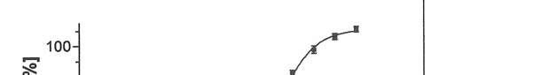
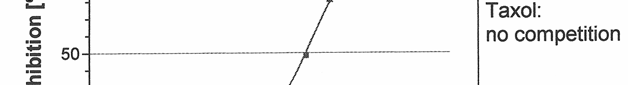

 Figure 1 - Detection of biotin tubulin and biological activity